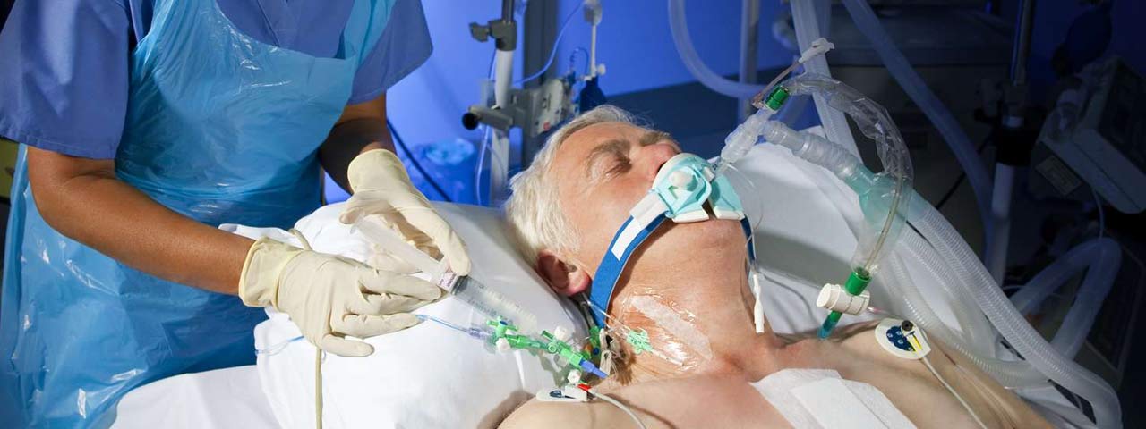
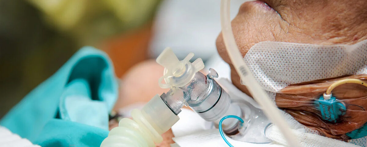
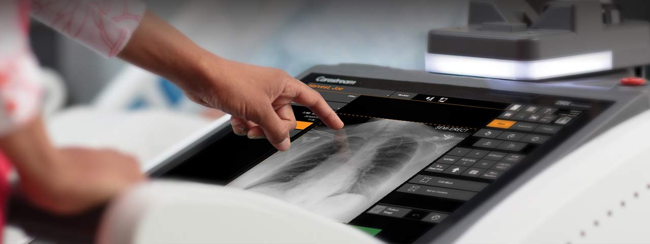
Digital Portable X-Ray is a diagnostic imaging technique that uses portable X-ray equipment to capture images of the body, such as the chest, abdomen, or extremities, at the patient’s bedside or in remote locations. It is commonly used in emergency rooms, intensive care units (ICUs), and for patients who cannot be easily transported.
Key Data Captured in Digital Portable X-Ray Reports
1. Patient Information:
- Name
- Age
- Gender
- Medical record number
- Date and time of the X-ray
- Indication/reason for the X-ray (e.g., suspected pneumonia, fracture, foreign body).
2. Imaging Details:
- Body part imaged (e.g., chest, abdomen, extremity).
- Views captured (e.g., AP [anteroposterior], PA [posteroanterior], lateral).
- Image quality (e.g., adequate, suboptimal due to positioning or motion).
3. Radiological Findings:
- Observations (e.g., presence of fracture, opacity, fluid levels, abnormal gas patterns).
- Specific conditions detected:
- Chest X-ray: Pneumonia, pleural effusion, pneumothorax, cardiomegaly, lung nodules.
- Abdominal X-ray: Bowel obstruction, perforation, abnormal gas patterns.
- Skeletal X-ray: Fractures, dislocations, bone lesions.
- Foreign objects (e.g., medical devices like pacemakers, endotracheal tubes, or central lines).
4. Measurements (if applicable):
- Heart size (cardiothoracic ratio) on chest X-rays.
- Dimensions of abnormalities (e.g., size of a lesion or nodule).
5. Comparison (if applicable):
- Comparison to prior X-rays (e.g., “Improvement in consolidation,” “No significant change in nodule size”).
6. Impression/Conclusion:
- Summary of findings (e.g., “No acute abnormalities,” “Findings suggest pneumonia”).
- Recommendations for further imaging or follow-up (e.g., CT scan for further evaluation).
Data Extraction from Reports
If extracting data from multiple Digital Portable X-Ray reports, organize it into a table:
| Patient Name | Age | Date | Body Part | View | Findings | Impression | Recommendations |
| John Doe | 45 | 2024-12-11 | Chest | AP | Opacity in left lower lobe | Suggestive of pneumonia | Follow-up with antibiotics |
| Jane Smith | 60 | 2024-12-10 | Left Wrist | Lateral | Fracture of distal radius | Confirmed distal fracture | Apply cast, X-ray in 2 wks |
Let me know if you have specific files or data to process, and I can help extract the relevant information and organize it into a report!

